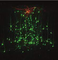
Single-cell-initiated monosynaptic tracing reveals layer-specific cortical network modules.
Wertz A*, Trenholm S*, Yonehara K, Hillier D, Raics Z, Leinweber M, Szalay G, Ghanem A, Keller G, Rózsa B, Conzelmann KK, Roska B (2015)
Science 349: 70-74. *equally contributed. [PDF]
We developed a technique for transsynaptic labeling from single neurons in layer 2/3 of the visual cortex and identified layer-specific structures hidden in the orientation-selective circuit network. Yonehara contributed by creating (almost) all of the plasmids and viruses used in the experiments.
The first stage of cardinal direction selectivity is localized to the dendrites of retinal ganglion cells.
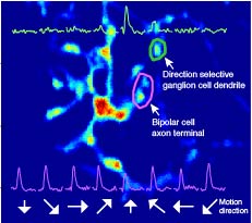
Yonehara K*, Farrow K*, Ghanem A, Hillier D, Balint K, Teixeira M, Jüttner J, Noda M, Neve RL, Conzelmann KK, Roska B (2013)
Neuron79: 1078-1085.*equally contributed.[ PDF]
Combining trans-synaptic labeling with GCaMP-expressing rabies virus and two-photon calcium imaging, we have developed a method to record synaptic activity in a monosynaptic network in a single network. The rabies virus development was done in collaboration with Dr. Karl-Klaus Conzelmann at LMU Munich.
Motion detection: Neuronal circuit meets theory.
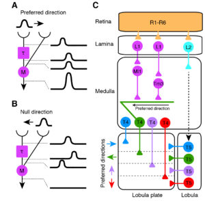
Yonehara K, Roska B (2013)
Cell 154:1188-1189. [ PDF]
Preview paper. A brief summary of recent findings on the structure and function of visuomotor processing circuits in Drosophila.
Spatially asymmetric reorganization of inhibition establishes a motion-sensitive circuit.
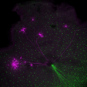
Yonehara K,Balint K, Noda M, Nagel G, Bamberg E, Roska B (2011)
Nature 469: 407-410. [ PDF]
Asymmetric inhibitory connections from starburst cells to SPIG1-positive directionally selective cells develop in a visual experience-independent manner between 6 and 8 days of age, as revealed by developmental optogenetics circuit mapping and trans-synaptic labeling with genetically modified rabies virus. We used the then-current, highly sensitive ChR2 developed by Drs. Bamberg and Nagel.
Identification of retinal ganglion cells and their projections involved in central transmission of information about upward and downward image motion.
Yonehara K, Ishikane H, Sakuta H, Shintani T, Nakamura-Yonehara K, Kamiji NL, Usui S, Noda M (2009)
PLoS One 4: e4320. [PDF]
We found that the mosaic of ganglion cells labeled by SPIG1 are directionally selective cells with upward-motion selectivity. The electrophysiological recording was done by Dr. Hiroshi Ishikane et al.
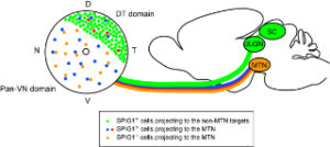
Expression of SPIG1 reveals development of a retinal ganglion cell subtype projecting to the medial terminal nucleus in the mouse.
Yonehara K, Shintani T, Suzuki R, Sakuta H, Takeuchi Y, Nakamura-Yonehara K, Noda M (2008)
PLoS One 3: e1533. [ PDF]
This is the first demonstration of genetic labeling of a single type of retinal ganglion cell that projects to the accessory optic system using the SPIG1 promoter. This work clarified the structure and development of this cell type in detail. This work opened up a new field of cell-type biology of retinal ganglion cells. This was Yonehara’s dissertation.